The Pterygoid Process
The pterygoid process is a paired bony structure projecting downward from the junction of the body and the greater wing of the sphenoid bone. It sits directly behind the maxillary tuberosity at the back of the upper jaw.
The pterygoid process (Latin: processus pterygoideus ossis sphenoidalis, from Greek pteron, wing) is a bony projection that descends from the base of the sphenoid bone at the back of the skull. In the context of dental implantation, it is one of the most important anatomical structures available for posterior maxillary anchorage. It does not resorb after tooth loss. It is connected to the skull base. And for patients with severe upper jaw bone loss who have been told they need sinus lift surgery or zygomatic implants, the pterygoid process can be the reason why they need neither.
Each pterygoid process consists of two plates:
- The medial pterygoid plate (Latin: lamina medialis processus pterygoidei) is the inner plate. It is the primary cortical anchorage target for tubero-pterygoid implants. It forms the lateral wall of the nasal cavity at its lower end and articulates with the palatine bone.
- The lateral pterygoid plate (Latin: lamina lateralis processus pterygoidei) is the outer plate. It provides attachment for the lateral pterygoid muscle, which is involved in jaw movement.
Between and around these plates, several important muscles attach, including the medial and lateral pterygoid muscles and the tensor veli palatini. For implant planning, the key anatomical focus is on the cortical bone of the medial plate and the junction between the pterygoid process and the maxillary tuberosity.
The tuberosity-pterygoid junction is the area where the posterior surface of the maxillary tuberosity meets the pterygoid process. This junction contains dense cortical bone on both sides. In strategic implantology, it is a defined anatomical target for distal maxillary implant anchorage.
The Bone Quality of the Pterygoid Process
Is the pterygoid bone dense enough to hold an implant?
Yes. The cortical bone of the pterygoid process is dense and mechanically reliable. Unlike the soft cancellous alveolar bone of the posterior maxilla, which resorbs progressively after tooth loss, the pterygoid process is connected to the base of the skull and does not undergo the same resorption process.
In terms of bone type classification, the pterygoid bone is typically D1 to D2 cortical bone. The medial pterygoid plate in particular contains compact, well-mineralized cortical bone that does not depend on the presence of teeth or alveolar bone to maintain its structure.
This is the critical clinical distinction. A patient who has lost most of the cancellous alveolar bone in the posterior maxilla may have very soft D4 bone in the remaining ridge. But at the pterygoid process, the bone quality is entirely different. The local alveolar bone is D4 while the anchorage target, the pterygoid cortex, is closer to D1 or D2. A correctly designed implant can reach from the depleted alveolar zone through the tuberosity-pterygoid junction and into the medial plate, bypassing the problem bone entirely.
The Pterygoid Process in Strategic Implantation
Why would my dentist plan an implant all the way back to the pterygoid process?
The posterior maxilla is the anatomical zone most commonly excluded from conventional implant treatment in patients with bone loss. The maxillary sinuses occupy the space above the molar region. After tooth loss, the sinus floor often drops further, leaving only a thin layer of bone above it. Conventional implants placed into this thin layer have poor primary stability and a high risk of failure.
The traditional answer to this problem is a sinus lift procedure, where the sinus membrane is raised surgically and bone graft material is packed below it to create enough height for conventional implants. This adds months to treatment, carries a significant failure risk, especially in patients whose bone is already compromised by gum disease, and does nothing to restore molar function at the back of the arch.
Pterygoid anchorage offers an anatomical alternative. By directing an implant at an oblique posterior angle from the tuberosity-pterygoid area into the medial plate of the pterygoid process, the surgeon completely bypasses the sinus. The implant anchors in bone that is dense, stable and not dependent on the quality of the depleted alveolar ridge.
For the patient, this means:
- No sinus lift
- No bone graft
- Distal molar support that allows full chewing function
- Immediate loading when primary stability is confirmed
Planning this accurately requires CBCT imaging to map the exact position of the medial plate, the distance from the tuberosity junction, the angulation needed and the implant length required to reach the target cortical structure safely.
Implant Selection for Pterygoid Process Anchorage
Which implants are designed to reach the pterygoid process?
Not every implant system is designed for pterygoid anchorage. The implants used in this zone must have sufficient length and the right thread geometry to engage cortical bone at depth, along with a bendable or angulated abutment to compensate for the oblique insertion angle.
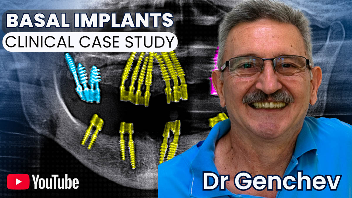
Ihde Dental BCS is one of the most widely documented implant systems for pterygoid anchorage. In Dr Genchev’s edentulism case study, a BCS implant at 3.5 x 20 mm was placed at position 17-2 at a posterior oblique angle specifically to reach the medial plate of the pterygoid process of the sphenoid bone. This provided the primary distal tubero-pterygoid anchor for the upper bridge. A second BCS implant at position 17 was placed through the tuberosity-pterygoid junction for additional posterior support.
Ihde Dental TPG Uno is also used in the pterygoid region when both cancellous and cortical bone are present in the tuberosity-pterygoid area. In the same case, Dr Genchev placed three TPG Uno implants in the left posterior maxilla at positions 25, 26 and 27. The longest, at 3.5 x 17 mm at position 27, was used to reach deep cortical anchorage in the pterygoid plate. At position 25, the TPG Uno was attached toward the pyramidal process of the palatine bone, which articulates with the pterygoid plates. In Dr Genchev’s gum disease case study, one TPG Uno was placed at position 17 because cancellous bone remained and the implant was anchored into the tubero-pterygoid process.
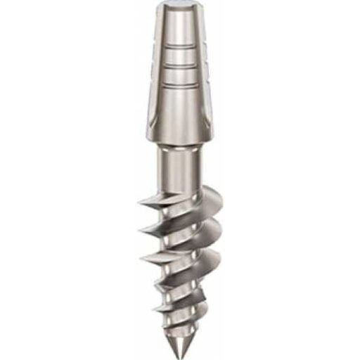
BasalFix TPI (Tubero-Pterygoid Implant) is an implant series specifically engineered for this zone. It is inserted at a posterior oblique angle and its target is the pterygoid process and tuberosity-pterygoid junction. The abutment can be bent up to 15 degrees to compensate for the insertion angulation. Typical dimensions for primary tubero-pterygoid anchorage are 3.5 x 20 mm at the most distal position, with wider dimensions such as 4.6 x 14 mm used through the tuberosity-pterygoid junction. The BasalFix TPI replaces the need for a sinus lift with a single oblique cortical fixation behind the sinus.
The BasalFix Basal can also be used in the pterygoid zone as a pure cortical anchorage implant where the anatomy supports it.
Case Studies About The Pterygoid process
-
Basal Implants for Edentulism and Bone Atrophy
Clinical case study of Dr Genchev’s basal implant protocol for total edentulism and severe bone atrophy
-
Dr Genchev Dental Restoration for Gum Disease
Dr. Genchev transformed Véronique’s life in just 5 days with basal implants, restoring her ability to eat without pain and giving her back a confident smile.
-
Dental restoration for missing teeth and bone atrophy with basal implants
Dr Genchev completed the full mouth dental restoration for this patient who suffered from edentulism with severe bone atrophy in 1 visit of 5 days. The price for the full dental restoration was 8990 Euro.
