The maxilla (Latin: maxilla, from mala, jaw) is the upper jaw. It is a paired bone that forms the central part of the facial skeleton, housing the upper teeth and forming the floor of the nasal cavity, the orbital floor and the roof of the mouth. For implantology, the maxilla is the more challenging of the two jaws because its bone is generally less dense than the mandible, and it contains structures such as the maxillary sinuses that limit where implants can be placed.
The maxilla consists of two fused bones that together form the upper jaw. For implant purposes, it is useful to think of it in three zones:
The anterior maxilla is the front section, roughly from the central incisors to the first premolars. It contains the premaxilla (Latin: praemaxilla), also called the intermaxillary bone, which carries the four upper incisors. The bone here tends to be D3 type: thin cortical shell, finer trabecular structure, less mechanical resistance than the mandible.
The mid-maxilla is the transitional zone between the anterior and posterior regions. Bone quality and volume can vary significantly here depending on the patient’s age, history of tooth loss and degree of resorption.
The posterior maxilla is the most challenging zone. It contains the molar region, the maxillary tuberosity (Latin: tuber maxillae) and lies immediately below and around the maxillary sinuses. In many patients, particularly those who have been edentulous for some time, the alveolar bone in this zone has resorbed, the sinus floor has dropped and the available bone for conventional implants is very limited.
The alveolar process (Latin: processus alveolaris) of the maxilla is the tooth-bearing ridge. Like the mandible’s alveolar process, it resorbs after tooth loss. But the deeper structures of the maxilla, including the cortical nasal floor, the palatine bone and the pterygoid process behind the tuberosity, do not resorb. These are the structures that basal implantology targets.
The Bone Quality of the Maxilla
Is my upper jaw bone good enough for implants?
Bone quality in the maxilla varies considerably by zone and is typically classified using the D1 to D4 Misch scale.
Anterior maxilla bone is most commonly D3. This means a thin cortical layer with fine trabecular bone underneath. It is not as mechanically forgiving as D1 or D2. Primary stability must be achieved carefully, often using controlled cancellous compression and the nasal floor cortex as a secondary anchorage point.
Posterior maxilla bone is often D4, particularly after significant tooth loss. D4 bone is very soft, fine trabecular bone with limited cortical support. The local alveolar bone in the posterior maxilla may be reduced to the point where conventional implant placement is impossible without sinus lift or bone grafting. However, the deeper cortical structures behind the tuberosity, including the pterygoid process, the palatine bone and the tuberosity-pterygoid junction, are not subject to resorption and remain dense and mechanically reliable. These structures are what make tubero-pterygoid implantation possible.
The key clinical insight is this: while the surface bone of the posterior maxilla is soft and shrinking, the anchoring structures behind and below it remain dense. The job of the implantologist is to reach those structures safely.
Anatomical Structures for Strategic Implantation
What anatomical landmarks does my dentist need to map before treating my upper jaw?
Several structures in the maxilla directly affect implant planning.
The maxillary sinuses (Latin: sinus maxillares, also called the antrum of Highmore) are air-filled cavities inside the maxilla on each side of the nose. They are the primary reason why conventional implant placement in the posterior upper jaw often requires sinus lift surgery. Basal implant techniques are specifically designed to work around the sinuses rather than through them.
The nasal floor (Latin: pars nasalis ossis frontalis) is the bony floor of the nasal cavity. In the anterior maxilla, this cortical structure lies above the apices of the upper incisor and canine roots. Implants placed in the anterior maxilla with sufficient length and slight palatal inclination can engage this cortical layer, providing a reliable secondary anchorage point even when the alveolar ridge is narrow or reduced.
The palatal cortical plate (Latin: lamina horizontalis ossis palatini) is the cortical bone forming the roof of the mouth. In the anterior and mid-maxilla, palatal inclination of implants can engage this cortical structure and improve primary stability.
The maxillary tuberosity (Latin: tuber maxillae) is the rounded prominence at the posterior end of the maxilla. Its cortical junction with the pterygoid process of the sphenoid bone is a key anchorage zone for distal maxillary implants.
The pterygoid process of the sphenoid bone is the bony column behind the tuberosity. It connects to the base of the skull and does not resorb regardless of how much maxillary bone is lost. It is discussed in detail on the pterygoid process anatomy page.
The pyramidal process of the palatine bone is the small bony projection that articulates with the pterygoid plates of the sphenoid. It is part of the complex cortical anatomy behind the maxillary tuberosity that makes tubero-pterygoid anchorage possible.
Implant Selection for the Maxilla
Which implants are designed for the upper jaw?
Implant selection in the maxilla depends on which zone is being treated and what bone remains. A single patient often needs different implant types in different areas of the same upper arch.
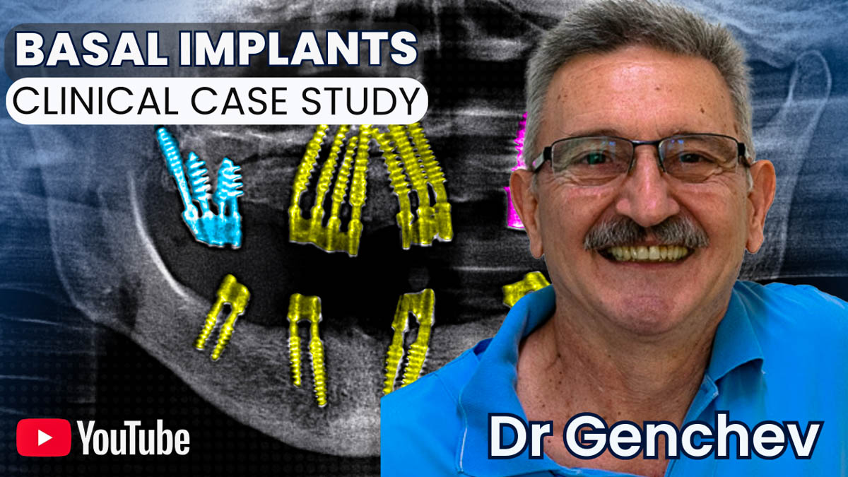
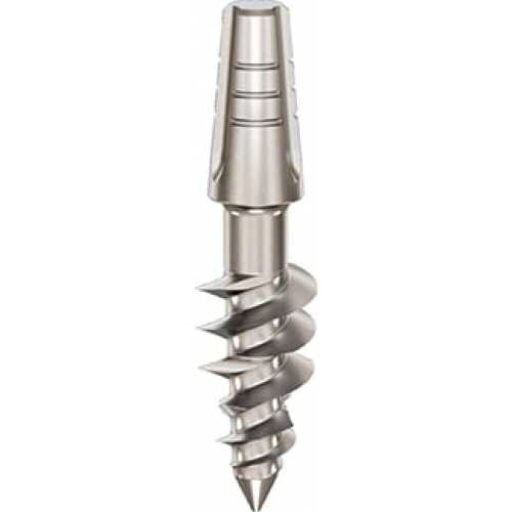
In the anterior maxilla, where D3 bone is common, the Ihde Dental KOS Root compression implant is often the preferred choice. It compresses the available cancellous bone during insertion and, when length and inclination allow, fixes its tip into the cortical bone of the nasal floor. In Dr Genchev’s case study of a patient with severe bilateral bone atrophy, he placed seven KOS Root implants at 3.5 x 16 mm across positions 14 to 23, each with slight palatal inclination, achieving cancellous compression and nasal floor cortical fixation simultaneously.
The Ihde Dental TPG Uno is also relevant in the anterior maxilla and mid-maxilla where both cancellous and cortical bone are available. Its triple-thread design allows it to engage the crestal cortical layer, compress cancellous bone and anchor in deeper cortical structures. In Dr Genchev’s gum disease case, TPG Uno implants were placed in the anterior maxilla to use both the cancellous bone and the cortical bone of the nasal floor.
In the posterior maxilla where cancellous bone has resorbed, the Ihde Dental BCS is used for pure cortical anchorage. In Dr Genchev’s case, BCS implants in the right posterior maxilla were placed into the tuberosity-pterygoid junction, the medial plate of the pterygoid process of the sphenoid, and the vertical process of the palatine bone. Where both cancellous and cortical bone remained, as in the left posterior maxilla of the same patient, TPG Uno was selected to combine compression with cortical fixation.
The BasalFix TPI (Tubero-Pterygoid Implant) is specifically engineered for the posterior upper jaw. It anchors into the pterygoid process and tuberosity-pterygoid junction, bypassing the sinus entirely without requiring a sinus lift or bone graft.
The BasalFix Compressive-Fix is useful in transition zones of the anterior to mid-maxilla, where the nasal cortex is reachable and some cancellous bone remains. It combines cancellous compression with cortical tip engagement.
For soft D3 and D4 maxillary bone, Monoimplant Rough fixtures support biological osseointegration through their calcium phosphate treated surface. The Monoimplant MOT, a hybrid with a rough upper surface and polished apical tip, is relevant near the sinus because its polished tip reduces contamination risk in that region.
Case Studies For Basal Dental Implants
-
Basal Implants for Edentulism and Bone Atrophy
Clinical case study of Dr Genchev’s basal implant protocol for total edentulism and severe bone atrophy