In dental implantology, implant length and diameter as well as its insertion angle and loading protocol, flow from the anatomical assessment made before surgery begins.
The Mandible
The lower jaw as an implant foundation
The mandible (lower jaw) is the only moveable bone of the skull. It generally has the best bone quality of the two jaws. Its anterior zone is associated with D1 and D2 bone: dense, well-mineralized cortical bone with good vascularity. Its posterior zones present more variability but even in atrophied lower jaws, the cortical plates often remain present and usable.
The mandible is the most reliable implant foundation in the dentition. Its anterior zone (symphysis and parasymphysis) contains D1 cortical bone, among the densest bone in the body. The interforaminal zone between the two mental foramina is particularly safe for implant placement because the inferior alveolar nerve exits through the mental foramina on each side, leaving the anterior arch free of major nerve risk.
The mandible carries two other structures that are directly relevant for posterior implant planning: the inferior alveolar nerve canal and the mylohyoid line. Planning mandibular implants requires CBCT imaging to map the nerve path precisely and to identify the cortical anatomy available for anchorage.
Bone type: Predominantly D1 (anterior) and D2 (body and premolar zone). Posterior mandible variable.
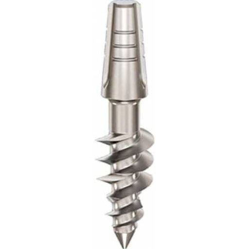
Dental implants: Ihde Dental KOS Root, TPG Uno, BCS; BasalFix Compressive, Compressive-Fix; Monoimplant Smooth, Microthread.
The Mylohyoid Line
Cortical anchorage in the posterior lower jaw
The mylohyoid line is a cortical ridge on the inner (lingual) surface of the mandibular body. It is the attachment site of the mylohyoid muscle and runs obliquely from the molar zone toward the midline. Its clinical importance is that it remains densely mineralized even when the alveolar ridge above it has substantially resorbed.
In patients with limited posterior mandibular bone height, a conventionally placed vertical implant may be unable to reach safe length without impinging on the inferior alveolar nerve. A lingually inclined implant can be directed past the nerve canal with its tip engaging the mylohyoid cortex. This converts an apparently impossible case into an achievable implant position.
In Dr Genchev’s edentulism case study, KOS Root implants in the posterior mandible were anchored into the mylohyoid line at positions 36, 37, 46 and 47, providing stable posterior support in bone that would have been inadequate for conventional vertical implant placement.
Bone type: D1 to D2 cortical ridge.
Dental implants: Ihde Dental KOS Root, BCS; BasalFix Compressive-Fix, Basal.
The Maxilla
The upper jaw and its anatomical zones
The maxilla (upper jaw) is anatomically more complex and more challenging. Its anterior zone has softer D3 bone: thinner cortical shell with finer trabecular structure. Its posterior zone is often D4: very soft cancellous bone, heavily influenced by the proximity of the maxillary sinuses. The posterior maxilla is the most common zone where conventional implants fail and where sinus lift procedures are recommended. Strategic basal implantology addresses this by targeting deeper cortical structures that do not resorb.
The maxilla demands zone-by-zone planning. No single implant type or strategy works across the entire upper arch. The anterior maxilla has D3 bone and offers narrow ridges that benefit from compression plus nasal floor cortex engagement. The posterior maxilla often presents D4 soft bone and sinus proximity. Its treatment requires access to the deeper cortical structures, including the tuberosity-pterygoid junction, the pterygoid process and the pyramidal process of the palatine bone.
A complete maxillary treatment plan in a patient with bone loss typically requires three or more different implant systems to address different bone conditions zone by zone. That is what experienced basal implantologists plan for.
Bone type: D3 anteriorly; D4 posteriorly; variable in the mid-maxilla.
Dental implants: Ihde Dental KOS Root (anterior), BCS and TPG Uno (posterior); BasalFix TPI, Compressive-Fix; Monoimplant Rough, MOT.
The Pterygoid Process
Distal maxillary anchorage without sinus lift
The pterygoid process is the bony projection descending from the sphenoid bone directly behind the maxillary tuberosity. It consists of a medial and lateral plate. Its medial plate is the primary cortical target for tubero-pterygoid implants. Because it is connected to the base of the skull, it does not resorb after tooth loss regardless of how much maxillary bone has been lost.
The tuberosity-pterygoid junction, where the posterior maxilla meets the pterygoid process, is one of the most important cortical anchorage targets in all of implantology. It allows the implantologist to provide distal posterior support for an upper bridge, restoring molar function, without sinus lift, bone graft or zygomatic implants.
In Dr Genchev’s edentulism case study, BCS implants reached the medial pterygoid plate of the sphenoid bone on the right side. TPG Uno implants engaged the pterygoid cortex on the left side. In his gum disease case, both BCS and TPG Uno implants were placed into the tubero-pterygoid process.
Bone type: D1 to D2 cortical bone (medial plate and tuberosity-pterygoid junction).
Dental implants: Ihde Dental BCS, TPG Uno; BasalFix TPI, Basal.
The Pyramidal Process
Supporting cortical anchorage in the mid-posterior maxilla
The pyramidal process is a compact bony projection of the palatine bone that fits into the notch between the pterygoid plates. It sits at the convergence of the palatine bone, the maxilla and the sphenoid. In practical terms, it is part of the same cortical complex as the pterygoid process and the palatine bone, and it offers an additional anchorage reference for mid-posterior maxillary implants.
It is smaller and more variable than the medial pterygoid plate and must be individually mapped using CBCT before it can be used as a planned cortical target. When it is accessible, it allows the implantologist to anchor an implant in a zone where alveolar bone has resorbed, using cortical bone that does not behave like the soft D4 tissue surrounding it.
In Dr Genchev’s edentulism case study, a TPG Uno implant at position 25 was specifically anchored toward the pyramidal process, forming part of the three-implant posterior left maxillary support.
Bone type: D1 to D2 compact cortical bone.
Dental implants: Ihde Dental TPG Uno, BCS; BasalFix TPI, Basal.
The Nasal Floor Cortex
Secondary cortical anchorage in the anterior maxilla
The nasal floor cortex is the dense bony floor of the nasal cavity. It lies immediately above the root apices of the upper incisors and canines. In the anterior maxilla, where D3 bone is the norm and alveolar ridge width is often limited, the nasal floor cortex provides a reliable secondary cortical anchor for implants placed with sufficient length and a slight palatal inclination.
The implant body compresses the cancellous bone in the alveolar process. The implant tip fixes into the cortical nasal floor above. This two-point anchorage significantly improves primary stability in a zone where cancellous compression alone would often be insufficient for immediate loading.
In Dr Genchev’s edentulism case study, seven KOS Root implants across the anterior maxilla (positions 14 to 23) were all placed with palatal inclination and all anchored their tips into the nasal floor cortex. This strategy made immediate loading of the full upper bridge possible in a zone with narrow bone width.
Bone type: D1 to D2 thin but dense cortical bone.
Dental implants: Ihde Dental KOS Root, TPG Uno; BasalFix Compressive-Fix, Basal.