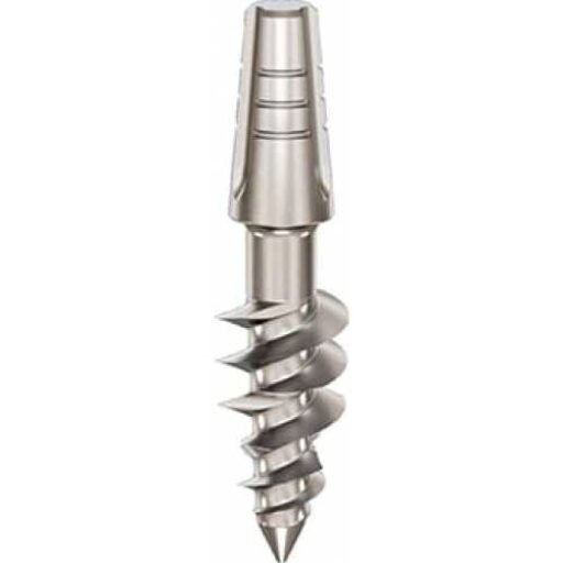
D4 bone is very soft, fine trabecular bone with little cortical support.
D4 bone is biologically vascular but mechanically weak. The challenge is achieving enough primary stability without relying on soft trabecular bone alone.
In clinical implant planning, D4 bone should not be treated as a simple label. It is a guide to how the bone may respond to drilling, compression, insertion torque and loading. The final diagnosis must always be based on CBCT assessment, clinical examination and tactile feedback during preparation.
Bone Location and Relevant Anatomy
D4 bone is most commonly associated with the posterior maxilla, especially the molar region, maxillary tuberosity and areas close to the maxillary sinus. The local alveolar bone may be soft and reduced after tooth loss, while deeper cortical structures such as the pterygoid process, tuberosity-pterygoid junction, palatine bone and sinus floor region may become important anchorage references.
Important anatomical references for D4 bone include:
- posterior maxilla
- maxillary molar region
- maxillary tuberosity
- maxillary sinus floor
- tuberosity-pterygoid junction
- pterygoid process of sphenoid bone
- vertical process of palatine bone
For implant treatment, these structures matter because they influence primary stability, drilling resistance, heat generation, prosthetic distribution and the possibility of strategic cortical anchorage.
Clinical Significance for Dental Implant Treatment
D4 bone is biologically vascular but mechanically weak. The challenge is achieving enough primary stability without relying on soft trabecular bone alone.
Drilling Protocol
Under-drilling is often necessary to improve primary stability, but it must be controlled. In severe posterior maxillary atrophy, the strategy may shift from trabecular retention to cortical or pterygoid anchorage.
Insertion Torque
Lower torque may be acceptable when the implants are distributed and protected. Stability must be assessed clinically; soft bone should not be forced to produce artificial high torque.
Healing Time and Loading Protocol
Delayed loading is often recommended in isolated conventional cases. Immediate loading may be possible only when primary stability is achieved through strategic cortical anchorage, adequate implant number, rigid splinting and carefully controlled occlusion.
Implant Design
Strong taper, deeper threads, aggressive thread design, rough surfaces or cortical/pterygoid anchorage designs may be needed, depending on the surgical concept.
Implant Types Relevant to D4 Bone
The implant type should be selected according to the bone available in each zone, not simply according to the name of the bone type. In advanced implantology, the same jaw may contain several different bone conditions, so a full-arch case may require several implant designs.
Ihde Dental BCS
BCS is relevant in posterior maxillary atrophy where the local cancellous bone is insufficient and the implant must engage stable cortical structures such as the tubero-pterygoid region or palatine bone.
Ihde Dental TPG Uno
TPG Uno is relevant when some cancellous bone remains but additional cortical anchorage is available, for example toward the pterygoid process or pyramidal process.
BasalFix TPI
BasalFix TPI is specifically relevant for posterior maxillary anchorage in the tubero-pterygoid region, bypassing the sinus and using dense cortical structures behind the maxilla.
BasalFix Basal
BasalFix Basal may be used in severe atrophy where the objective is engagement of the basal or second cortical plate rather than relying on soft cancellous bone.
Monoimplant Rough
Rough Monoimplant is described for soft D3 and D4 bone and is intended to support biological osseointegration.
Monoimplant MOT
MOT Monoimplant is relevant in selected maxillary sinus-area cases because of its hybrid rough and polished surface design.
Clinical Case Study: Dr Genchev and Severe Bone Atrophy
Marie’s posterior maxilla is the clearest clinical example for D4-type challenges. On the right side, cancellous bone had resorbed and Dr Genchev selected BCS implants for cortical fixation into the tubero-pterygoid region, medial pterygoid plate and vertical process of the palatine bone. On the left side, where both cancellous and cortical bone remained, he used TPG Uno implants to combine compression with cortical anchorage. This shows how soft posterior maxillary bone can be managed by shifting the objective from local trabecular stability to strategic cortical anchorage.
In the wider case, Dr Genchev treated a totally edentulous patient with severe bilateral bone atrophy. He selected different implant types according to the bone available in each zone: BCS for hard cortical anchorage, TPG Uno where cancellous and cortical bone were both available, and KOS Root where cancellous compression could provide primary stability. The case was restored with fixed PFM metal-ceramic bridges within five days.
Practical Summary
- D4 bone must be assessed with CBCT and clinical judgment.
- The drilling protocol should match the density and vascularity of the bone.
- Implant design should respect the biology of the bone, not only the desired torque value.
- Immediate loading depends on primary stability, implant distribution, prosthetic splinting and occlusal control.
- In atrophic jaws, strategic anatomical anchorage may be more important than the local density of the alveolar ridge alone.
FAQ – Questions about bone type D4
Where is D4 bone usually found?
D4 bone is most commonly found in the posterior maxilla, especially the molar and tuberosity areas near the maxillary sinus.
Does D4 bone always need grafting?
Not always. Some cases require grafting or sinus lift, but strategic implantology may use cortical or pterygoid anchorage when anatomy allows.
Which implants are relevant in D4 posterior maxilla?
Implants designed for soft bone or strategic cortical anchorage may be relevant, including Ihde Dental BCS, TPG Uno, BasalFix TPI, BasalFix Basal, Monoimplant Rough and Monoimplant MOT.