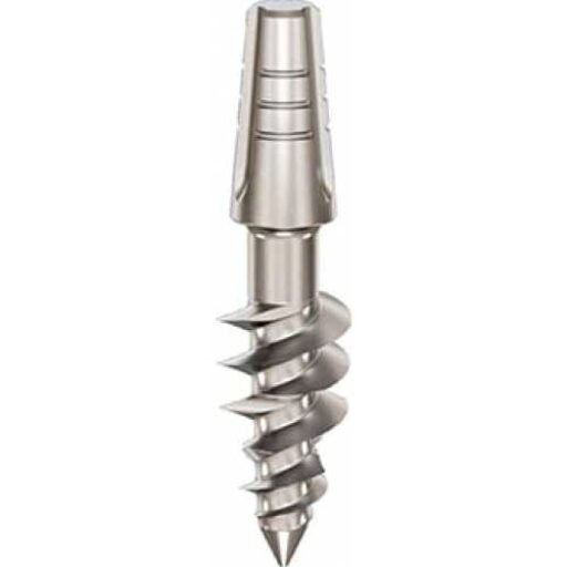
D2 bone is thick porous cortical bone with a strong trabecular inner structure.
D2 bone is often considered one of the most favorable conditions for implant placement because it combines good mechanical support with better vascularity than pure cortical bone.
In clinical implant planning, D2 bone should not be treated as a simple label. It is a guide to how the bone may respond to drilling, compression, insertion torque and loading. The final diagnosis must always be based on CBCT assessment, clinical examination and tactile feedback during preparation.
Bone Location
D2 bone is commonly found in the anterior mandible and in many mandibular body regions. It may also be present in parts of the anterior maxilla depending on the patient’s age, anatomy and degree of resorption.
Important anatomical references for D2 bone include:
- anterior mandible
- posterior mandible
- mandibular body
- premolar region
- coarse trabecular bone
- porous cortical bone
- alveolar process
For implant treatment, these structures matter because they influence primary stability, drilling resistance, heat generation, prosthetic distribution and the possibility of strategic cortical anchorage.
Clinical Significance for Dental Implant Treatment
D2 bone is often considered one of the most favorable conditions for implant placement because it combines good mechanical support with better vascularity than pure cortical bone.
Drilling Protocol
A standard drilling protocol is often appropriate, adapted to implant diameter, length and manufacturer protocol. The surgeon still controls heat, irrigation and pressure carefully.
Insertion Torque
D2 bone often supports strong primary stability. Torque targets must remain biological, not purely mechanical, because over-compression can still injure living bone.
Healing Time and Loading Protocol
D2 bone often allows flexible loading decisions. Immediate loading may be considered if primary stability, implant distribution and rigid prosthetic splinting are adequate.
Implant Design
Tapered or hybrid implants often work well. Compression implants can be useful when cancellous bone is present and can be condensed safely.
Implant Types Relevant to D2 Bone
The implant type should be selected according to the bone available in each zone, not simply according to the name of the bone type. In advanced implantology, the same jaw may contain several different bone conditions, so a full-arch case may require several implant designs.
Ihde Dental KOS Root
KOS Root is relevant where cancellous bone is present and can be compressed to create primary stability. In D2 bone this may provide a favorable mechanical and biological balance.
Ihde Dental TPG Uno
TPG Uno is relevant where the implant can use both cancellous compression and cortical engagement, especially in splinted fixed bridge work.
BasalFix Compressive
The BasalFix Compressive line is designed for zones with healthy cancellous bone where lateral compression can create immediate friction-based primary stability.
BasalFix Compressive-Fix
Compressive-Fix may be useful when D2 bone transitions toward a thinner or less dense zone and the clinician wants compression plus cortical tip engagement.
Monoimplant Smooth
Smooth Monoimplant is described for hard D1 and D2 bone and may be relevant when bicortical fixation and immediate loading are planned.
Clinical Case Study: Dr Genchev and Severe Bone Atrophy
In Marie’s lower anterior mandible, Dr Genchev used KOS Root implants in cancellous bone zones, adapting the length to local bone height. This is clinically relevant to D2 bone because KOS Root uses cancellous compression to obtain primary stability when the trabecular layer is present and strong enough.
In the wider case, Dr Genchev treated a totally edentulous patient with severe bilateral bone atrophy. He selected different implant types according to the bone available in each zone: BCS for hard cortical anchorage, TPG Uno where cancellous and cortical bone were both available, and KOS Root where cancellous compression could provide primary stability. The case was restored with fixed PFM metal-ceramic bridges within five days.
Practical Summary
- D2 bone must be assessed with CBCT and clinical judgment.
- The drilling protocol should match the density and vascularity of the bone.
- Implant design should respect the biology of the bone, not only the desired torque value.
- Immediate loading depends on primary stability, implant distribution, prosthetic splinting and occlusal control.
- In atrophic jaws, strategic anatomical anchorage may be more important than the local density of the alveolar ridge alone.
FAQ – Questions for D2 Bone Quality
Why is D2 bone favorable for implants?
D2 bone offers a good combination of cortical strength and trabecular blood supply, which helps both primary stability and healing.
Where is D2 bone found?
It is commonly found in the anterior mandible and in many areas of the mandibular body, with variation between patients.
Which implants are relevant for D2 bone?
Compression and hybrid implants may be relevant, including Ihde Dental KOS Root, TPG Uno, BasalFix Compressive and selected Monoimplant designs.