D3 bone is a thin cortical layer with fine trabecular bone.
D3 bone can support implants, but it is more technique-sensitive. It has less cortical support than D1 or D2 bone, so the surgeon must create primary stability without over-enlarging the osteotomy or damaging the fine trabecular network.
In clinical implant planning, D3 bone should not be treated as a simple label. It is a guide to how the bone may respond to drilling, compression, insertion torque and loading. The final diagnosis must always be based on CBCT assessment, clinical examination and tactile feedback during preparation.
Bone Location
D3 bone is often associated with the anterior maxilla, especially the premaxilla and anterior maxillary alveolar process. It may also be encountered in the posterior mandible in some patients.
Important anatomical references for D3 bone include:
- anterior maxilla
- premaxilla
- maxillary alveolar process
- nasal floor cortex
- palatal cortical plate
- posterior mandible
- fine trabecular bone
For implant treatment, these structures matter because they influence primary stability, drilling resistance, heat generation, prosthetic distribution and the possibility of strategic cortical anchorage.
Clinical Significance for Dental Implant Treatment
D3 bone can support implants, but it is more technique-sensitive. It has less cortical support than D1 or D2 bone, so the surgeon must create primary stability without over-enlarging the osteotomy or damaging the fine trabecular network.
Drilling Protocol
Controlled under-drilling may be useful, depending on the implant system. The preparation should preserve trabecular bone and allow the implant design to engage or compress it gently.
Insertion Torque
Moderate insertion torque is often sufficient. The clinician should avoid chasing high torque numbers if that would damage the bone.
Healing Time and Loading Protocol
Delayed loading is often preferred for conventional protocols. Immediate loading may be possible in selected full-arch or splinted cases if stability is distributed across several implants.
Implant Design
Deeper threads, tapered bodies, compression designs and hybrid compression-cortical fixation designs may improve stability in D3 bone.
Implant Types Relevant to D3 Bone
The implant type should be selected according to the bone available in each zone, not simply according to the name of the bone type. In advanced implantology, the same jaw may contain several different bone conditions, so a full-arch case may require several implant designs.
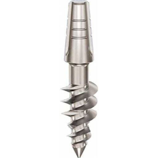
Ihde Dental KOS Root
KOS Root is relevant when cancellous bone is present and can be compressed. In the anterior maxilla it may also gain additional fixation if the tip reaches the nasal floor cortex.
Ihde Dental TPG Uno
TPG Uno is relevant in mixed cancellous and cortical situations where the body compresses cancellous bone while the apical area engages cortical bone.
BasalFix Compressive
BasalFix Compressive may be used where cancellous bone is still healthy enough to generate friction-based primary stability.
BasalFix Compressive-Fix
Compressive-Fix is especially relevant in transition zones, such as the anterior to mid-maxilla, where cancellous bone is present and the nasal floor cortex may be reachable.
Monoimplant Rough
Rough Monoimplant is described as suitable for softer D3 and D4 bone, with a surface designed to encourage osseointegration.
Monoimplant MOT
MOT Monoimplant is relevant in selected maxillary or sinus-related situations because it combines a rough upper portion with a polished apical tip.
Clinical Case Study: Dr Genchev and Severe Bone Atrophy
In Marie’s anterior maxilla, Dr Genchev used KOS Root implants with slight palatal inclination. The implants compressed the available cancellous bone and fixed their tips into the nasal floor cortex. This is highly relevant to D3 bone because the anterior maxilla often has a thinner cortical layer and finer trabecular bone, requiring both careful compression and a reliable cortical anchorage target.
In the wider case, Dr Genchev treated a totally edentulous patient with severe bilateral bone atrophy. He selected different implant types according to the bone available in each zone: BCS for hard cortical anchorage, TPG Uno where cancellous and cortical bone were both available, and KOS Root where cancellous compression could provide primary stability. The case was restored with fixed PFM metal-ceramic bridges within five days.
Practical Summary
- D3 bone must be assessed with CBCT and clinical judgment.
- The drilling protocol should match the density and vascularity of the bone.
- Implant design should respect the biology of the bone, not only the desired torque value.
- Immediate loading depends on primary stability, implant distribution, prosthetic splinting and occlusal control.
- In atrophic jaws, strategic anatomical anchorage may be more important than the local density of the alveolar ridge alone.
FAQ – Questions on D3 bone type
Where is D3 bone usually found?
D3 bone is often found in the anterior maxilla and may also occur in the posterior mandible.
Can D3 bone support immediate loading?
It can in selected cases, especially when implants are well distributed and rigidly splinted. In many conventional cases, delayed loading is safer.
Which anatomical structure is important in anterior maxillary D3 bone?
The nasal floor cortex can be an important secondary cortical anchorage target when implant length and angulation allow it.